Adipocytes Can Be Demostrated by Which of Th Following Techniques
ADSCs adipogenic differentiation is regulated by multiple. The adipocytes by screening into the desired size ranges.
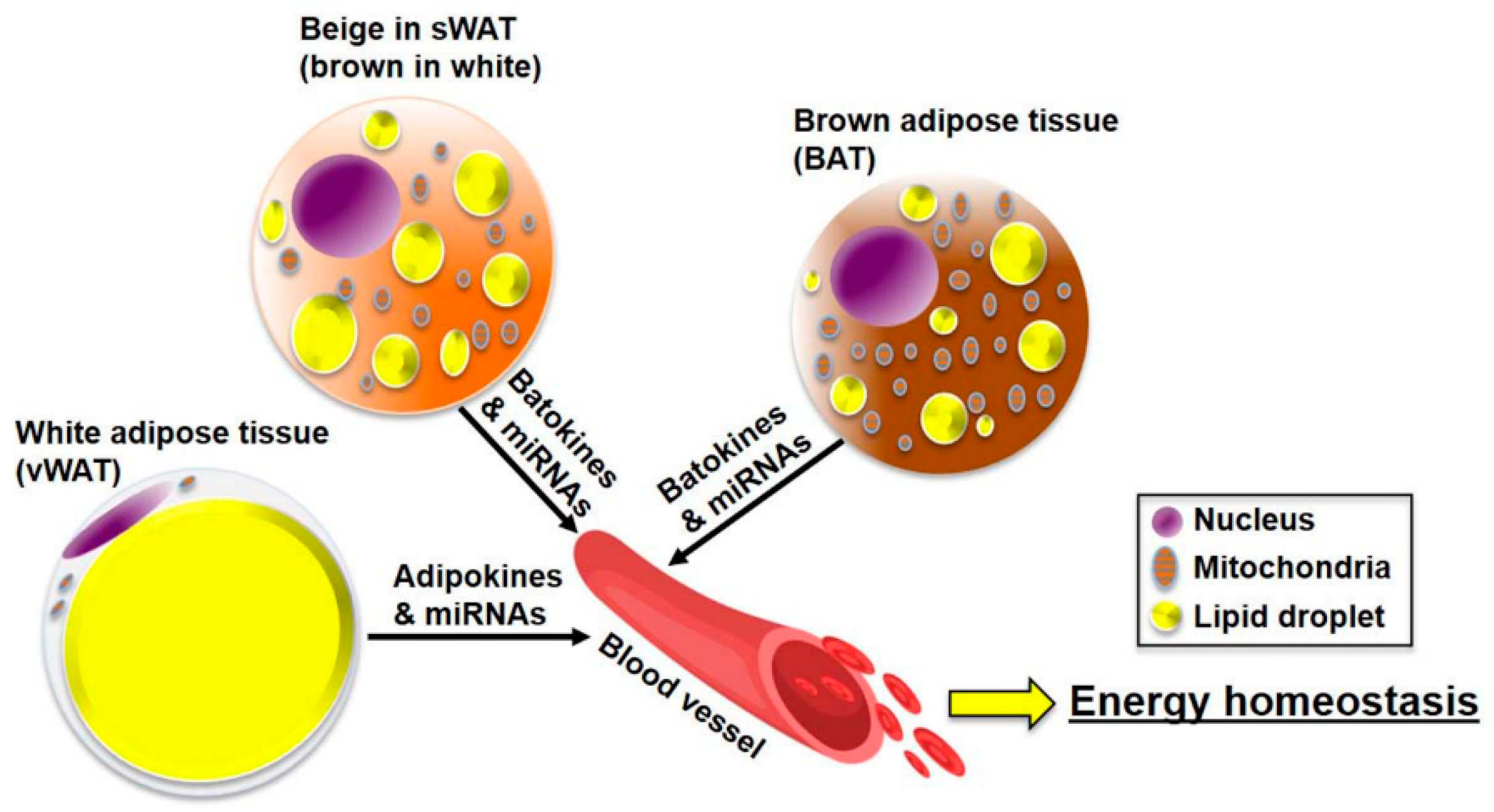
Jcm Free Full Text Adipose Tissue Derived Signatures For Obesity And Type 2 Diabetes Adipokines Batokines And Micrornas Html
This methodology directly improves previously published protocols can be utilized to study spatial distribution of other cell types within adipose tissue and will be a useful tool for investigating adipose tissue.

. 1999 and in vivo. Adipocyte apoptosis is the primary causes of fat graft volume loss resulted in variable absorption rate. 3 slides can be stained directly with picro-Ponceau.
Nutritional and pharmacological stimuli can dramatically alter the cellular phenotypes in white adipose tissue WAT. To increase following norepinephrine stimulation of. This inflammation can be connected with local loading of the reticular dermis with lipids released due to de-differentiation of adipocytes.
The sympathetic nervous system SNS plays a central role in the recruitment of brown adipocytes and its thermogenic activity through β3-AR stimulation 62. These costly techniques have focused on damaging adipocyte cell membranes hydrolyzing triglycerides TGs or inducing apoptosis. The optimal procedure for analysis of immature adipose depots consists of the following steps.
Adipocytes in various size fractions can then be counted sized and then decolorized with hydrogen peroxide in order to quantitate the amount of radioactivity within the adipocytes. Supporting this hypothesis it has been demonstrated that dedifferentiated adipocytes can be redifferentiated into adipocytes in vitro Kou et al. The main objective of this thesis is to demonstrate the potential of 13C-based techniques as tools for investigating metabolic rewiring in adipocytes.
Upon cold exposure free fatty acids FFAs released from intracellular lipid droplets or from TG-rich lipoproteins in the blood enter mitochondria and bind to UCP1 1. The following procedure describes our steps for staining free-floating adipocytes with the DNA stain DyeCycle Violet. There was no loss of radioactivity from the fixed cells with hydrogen peroxide treatment.
SSAT is located immediately beneath the skin and. 2 cryostat sections are cut removed from the knife with a room temperature slide and then air dried for 5-10 minutes. Utilizing viable adipose tissue as a free graft requires great care during the harvesting transfer and storage process for predictable results.
This report describes a novel method of adipocyte size and count measurement. 1 fresh unfixed tissues are rapidly in isopentane quenched in a liquid nitrogen bath. Cell Reports Report Transforming Growth Factor-b3 Regulates Adipocyte Number in Subcutaneous White Adipose Tissue Paul Petrus17 Niklas Mejhert17 Patricia Corrales2 Simon Lecoutre1 Qian Li1 Estela Maldonado3 Agne Kulyte1 Yamila Lopez2 Mark Campbell4 Juan R.
19 22 4245 Several studies have shown that the deep layer of the subcutaneous fat is the optimal site of harvest as it contains the highest concentration of mature adipocytes. Various fixatives as well as tissue and slide handling procedures have been evaluated in attempts to demonstrate adipocytes histochemically while maintaining cell and tissue integrity. Specifically we apply stable-isotope tracing methods and 13C-MFA to investigate metabolic rewiring in adipocytes exposed to three perturbations implicated in the pathogenesis of metabolic disorders.
Many techniques have been proposed for the harvesting of adipose tissue from a donor site before transfer including vacuum or syringe suction and surgical excision. Encoded water-fat MRI techniques that can separate. Adipocyte browning may better tolerate avascular environments and improve graft survival.
Mature adipocyte dedifferentiation increase the retention rate of fat grafts by acting as seed cells. SirT1 a potent inducer of adipose browning can significantly enhance the beige adipocyte differentiation capability in an elderly adipose-derived mesenchymal stem cell AD-MSC population. The optimal procedure for analysis of immature adipose depots consists of the following steps.
1 fresh unfixed tissues are frozen rapidly in isopentane quenched in a liquid nitrogen bath. This layer can also be further subdivided by the fascia superficialis which is visible by imaging techniques 6263 into superficial subcutaneous adipose tissue SSAT and deep subcutaneous adipose tissue DSAT. Utilizing genetic lineage tracing techniques we demonstrate that brown adipocytes BA that are induced by β3-adrenergic receptor activation in abdominal WAT arise from the proliferation and differentiation of cells expressing platelet-derived growth factor.
Nutritional and pharmacological stimuli can dramatically alter the cellular phenotypes in white adipose tissue WAT. Likewise basal adipocyte turnover is very low in rodents but can be accelerated by high-fat diet HFD feeding 91. Clinical outcomes following fat grafting are variable and technique dependent and it is unknown how the graft is revascularized.
Adipocytes from the superficial layer of subcutaneous adipose tissue undergo cyclic de- and re-differentiation which can significantly influence the development of skin inflammation under different cutaneous conditions. Utilizing genetic lineage tracing techniques we demonstrate that brown adipocytes BA that are induced by β3-adrenergic receptor activation in abdominal WAT arise from the proliferation and differentiation of cells expressing platelet. The study also demonstrated that the ratio.
The authors recently observed that. The series of histological samples here were only used to demonstrate the functionality and the consistency of this novel method as compared with an established alternative method ImageJ Adipocyte Tools with or without manual correction. Culture morphology was assessed by light microscopyACM cultures without 3T3s AT cultures with and without 3T3s and 3T3 control cultures demonstrated a similarly significant keratinocyte proliferation increase over non-3T3 control P 005 corresponding with a 2-fold increase in percent confluence by day 7.
Lineage tracing studies show that adipogenesis increases in visceral fat within 4 weeks of HFD feeding 93. The effect is depot-specific and higher in visceral versus subcutaneous fat 92. After incubating adipocytes with LipidTOX and fluorescent antibodies and washing the cells are resuspended to the previous volume and DyeCycle Violet is added to a dilution of 1333 to the suspension and mixed gently.
Viable adipocytes can be harvested via aspiration and yield successful soft-tissue augmentation in appropriate recipient sites. The canonical means of heat production in mouse and human adipocytes occurs through the mitochondrial transporter uncoupling protein 1 UCP1 1. Here we present a simple low-cost technique termed.
Associated Data Data Availability Statement. Acosta1 Jurga Laurencikiene1 Iyadh Douagi1 Hui Gao5 Concepcion Martınez-Alvarez3 Per. We have demonstrated the unique spatial distribution of mesothelium-derived Type-1 adipocytes in intact mesenteric fat using BABB-D4 clearing method.
The most superficial layer SAT is located beneath the skin and above the abdominal musculature.

Adipose Tissue Physiology To Metabolic Dysfunction Endotext Ncbi Bookshelf

Characteristics Of Adipocytes The Figure Represents Distinct Download Scientific Diagram
Adipose Tissue An Overview Sciencedirect Topics


Comments
Post a Comment